

INTRODUCTION
Gamma-delta T-cell acute lymphoblastic leukemia (γδ T-ALL) represents a distinct variant of lymphoblastic leukemia with unique clinical and biological features.1 Comprising approximately 10 to 15% of T-ALL cases, γδ T-ALL is uncommon and associated with a poor prognosis. In a study involving pediatric patients diagnosed with T-ALL, those expressing the γδ T-cell receptor displayed higher minimal residual disease (MRD) levels during remission induction and experienced lower overall survival rates compared to patients with alpha-beta (αβ) T-ALL.2
CASE REPORT
We report a case of a 16-year-old male who presented with ataxia, fever, fatigue, and evidence of encephalopathy. Physical exam demonstrated no appreciable hepatosplenomegaly, lymphadenopathy, gastrointestinal or skin involvement. Ultrasound revealed mild splenomegaly (18.9 cm in longest dimension). Their past medical history was insignificant with no history of chronic illnesses or previous admission to the hospital.
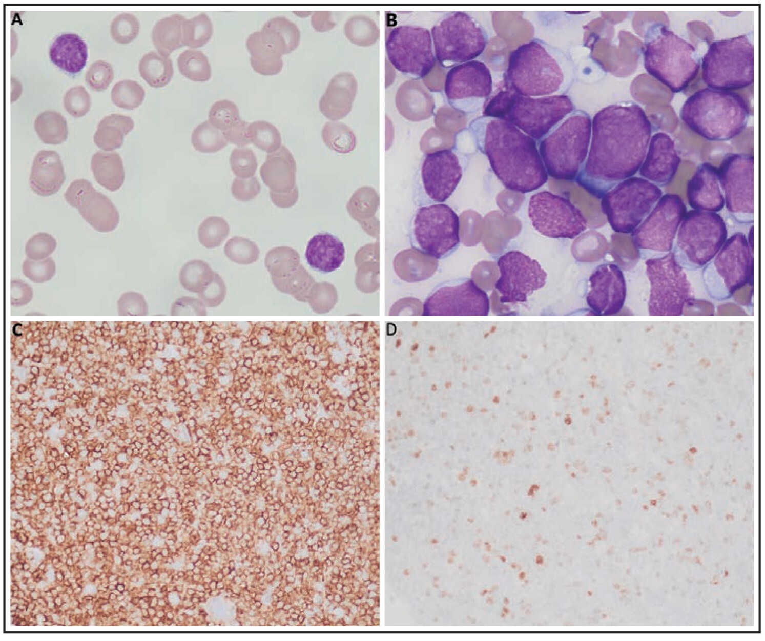
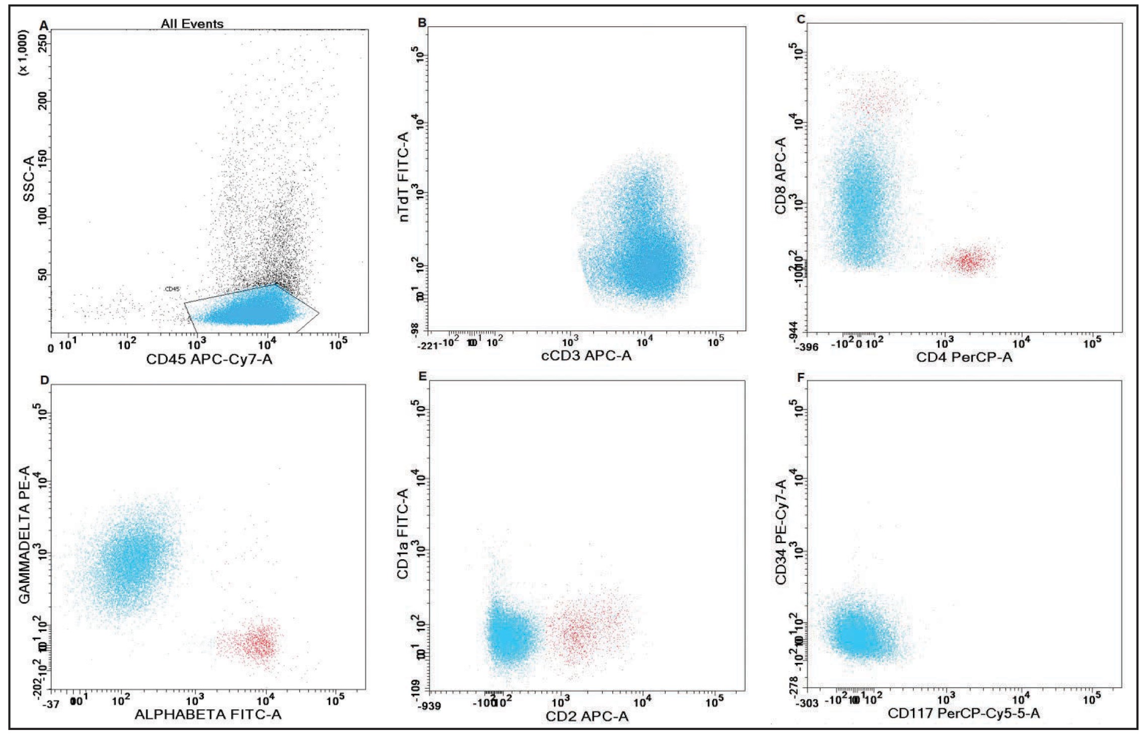
Their bloodwork showed pancytopenia with a hemoglobin of 83 g/L, thrombocytopenia of 35 x109/L, and mild leukocytosis of 12.77 x10⁹/L composed of approximately 90% abnormal lymphoid cells. Evidence of evolving transaminitis and disseminated intravascular coagulation (DIC) was noted, with the latter indicated by abnormalities in coagulation tests and the presence of occasional schistocytes observed by morphologic examination. The abnormal lymphoid population was predominantly comprised of small forms with variably dispersed chromatin and indistinct nucleoli (Figure 1A). Flow cytometric analysis of the peripheral blood revealed a population of neoplastic cells which were: positive for surface (s) and cytoplasmic (c) CD3, γδ T-cell receptor CD5, CD7 and CD52; variably positive for CD45; dimly positive for CD8; and negative for CD4, CD2, CD25, CD16, CD56, CD57, αβ T-cell receptor, CD1a, TdT, CD10, CD117, CD34, and B-cell and myeloid markers.
Bone marrow examination showed a markedly hypercellular marrow, packed with abnormal lymphoid cells that showed variability in size, mainly comprised of small to intermediate forms, and demonstrated blast morphology with dispersed chromatin, some with distinct nucleoli, and scant amount of a blue-gray agranular cytoplasm. The chromatin of the cells was considerably more delicate than what was observed on the peripheral blood (Figure 1B). Additional flow cytometric analysis of the bone marrow indicated that the neoplastic T-cells were negative for CD48 (determined by testing at a reference laboratory), while approximately 15% of cells tested positive for TdT, as confirmed by immunohistochemistry (IHC) (Figure 1D). The remaining markers displayed expression patterns consistent with those observed in the peripheral blood (Figure 2A-F). Notably, the cells exhibited strong positivity for CD99 by IHC (Figure 1C).
DISCUSSION
The differential diagnosis of a γδ T-cell neoplasm includes T-ALL, hepatosplenic T-cell lymphoma (HSTCL), T-cell large granular lymphocytic leukemia (T-LGL), and non-hepatosplenic nodal and extra nodal T-cell lymphomas.3 This case presented in a leukemic phase, with an aggressive clinical course and no lymph node, cutaneous, or GI involvement. Rare leukemic cases of HSTCL have been reported,4 however, the main sites of involvement are spleen (with significant splenomegaly contrary to this case), liver, and bone marrow. Furthermore, the IHCs for cytotoxic markers (TIA-1 and granzyme B) were negative in this case which further precludes a diagnosis of HSTCL.
It was difficult to definitively establish the stage of maturation of the abnormal T-cells by morphology and immunophenotype of the cells in the peripheral blood. However, morphology of the cells in the bone marrow was suggestive of blasts rather than mature T-cells. In addition, further immunophenotyping of the cells in the marrow confirmed their immature nature as they were strongly positive for CD99, with a subset of cells positive for TdT, while negative for CD48. CD99 is a nonspecific marker; however, in the setting of a T-cell neoplasm, it is mainly seen in T-lymphoblastic leukemia, anaplastic large cell lymphoma (ALCL), and peripheral T-cell lymphoma, not otherwise specified (PTCL, NOS).5,6 Regardless of other markers, ALCL was not a morphological concern in this case; additionally, leukemic presentation in PTCL, NOS is extremely rare, and most of the cases express αβ T-cell receptor.7,8 Reduced expression of CD48 is a marker for immaturity, differentiating mature T-cells from blasts in T-ALL, and is recognized as a useful marker to assess MRD in T-ALL after therapy.9–11
The diagnosis of γδ T-ALL was made based on the morphological characteristics of the neoplastic cells observed in the bone marrow and their immunophenotypic profile.
Involvement of the central nervous system was confirmed by morphologic examination (CNS3 positive status). Cytogenetic analysis identified MLLT4(AFDN)/MLL(KMT2A) fusion and a TP53 deletion (at17p13) in approximately 85% of nuclei. Additional FISH analysis to evaluate common chromosome abnormalities associated with specific T-cell lymphoma subtypes including the TCL1A gene region, chromosome 7, and chromosome 8 was within normal limits. Genetic alterations specific to the γδ T-cell lineage that result in fusion transcripts, such as SET-NUP214 and CALM-AF10, were not identified. SET-NUP214 and CALM-AF10 have been demonstrated to affect clinical outcomes and are implicated in chemotherapy resistance and poor prognosis in T-ALL patients.2,12,13
The patient underwent induction therapy as per Children’s Oncology Group (COG) protocol AALL1231.14 Subsequently, post-induction therapy was as per COG study protocol AALL0434, including the use of nelarabine.15 The patient is currently receiving maintenance chemotherapy consisting of oral mercaptopurine and weekly methotrexate, and remains in remission.
CONCLUSION
This report represents a case of a γδ T-ALL, a rare entity, with challenging morphologic findings in the peripheral blood that required extended immunophenotyping to reach final diagnosis. The clinical and pathologic findings underscore the complexity of diagnosing and managing such cases. Further studies are required to assess the prognostic significance of the identified genetic alterations and their potential therapeutic implications in γδ T-ALL.
This manuscript was peer reviewed.
Disclaimers
The authors declare that there are no undisclosed conflicts of interest regarding the publication of this paper. All authors have provided Clockwork Communications Inc. with non-exclusive rights to publish and otherwise deal with or make use of this article, and any photographs/images contained in it, in Canada and all other countries of the world.