INTRODUCTION
Perivascular epithelioid cell tumours (PEComas) are defined by the World Health Organization as “mesenchymal tumours composed of histologically and immunohistochemically distinctive perivascular epithelioid cells.”1 The PEComa family of neoplasms includes angiomyolipoma (AML), clear cell ‘sugar’ tumour of the lung, lymphangioleiomyomatosis (LAM), and a group of morphologically and immunophenotypically similar tumours arising elsewhere.2 PEComas have a strong female predominance and occur over a wide age range, with non-AML /non-LAM PEComas presenting at a median age of 46 years.3 Of the PEComas that arise in soft tissue, the retroperitoneum, viscera, abdominal wall, and pelvis are the most common locations.4 Primary PEComas of the bone are very rare, however over 20 such cases have been described in the literature to date.4–8 While the majority of PEComas demonstrate benign biological behavior, PEComas arising in bone and soft tissue sites have a greater propensity to metastasize than those arising elsewhere.5 The natural history of metastatic PEComas remains poorly understood due to their rarity. A systematic review of case reports found a median overall survival of 60 months in metastatic cases.9
Metastatic PEComas commonly harbour mutations that lead to mTOR pathway activation.10 For instance, recurrent biallelic inactivating molecular alterations in TSC1/TSC2 have been identified in approximately 42% of metastatic PEComas,11 including germline mutations associated with tuberous sclerosis complex.12 Recurring gene fusions involving TFE3 have also been identified in approximately 16% of cases.11 PEComas tend to also harbor a variety of additional mutations in genes such as TP53, ATRX, and MSH3, however mutations associated with mismatch repair deficiency (MMRd) and microsatellite instability (MSI) are rare.13
In solid tumours, MSI-high status has been shown to predict efficacy to immune checkpoint inhibition. MSI-high tumours typically have a high tumour mutation burden, which leads to increased neoantigen expression and increased tumour immunogenicity. These tumours show increased susceptibility to monoclonal antibodies that target programmed death receptor-1 (PD-1), thereby enhancing the immune system’s ability to recognize and destroy tumour cells. Here we report a case of MSI-H metastatic PEComa of bone origin that was successfully treated with pembrolizumab.
CASE PRESENTATION
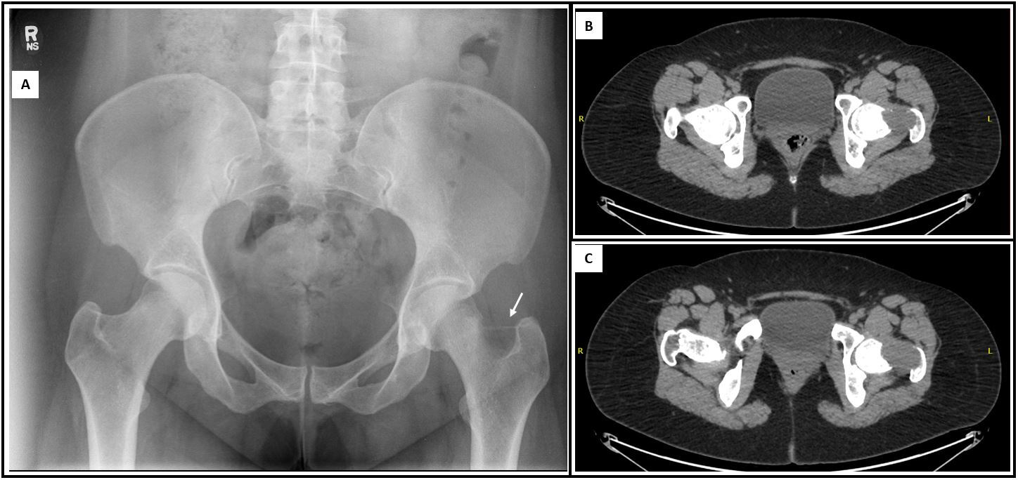
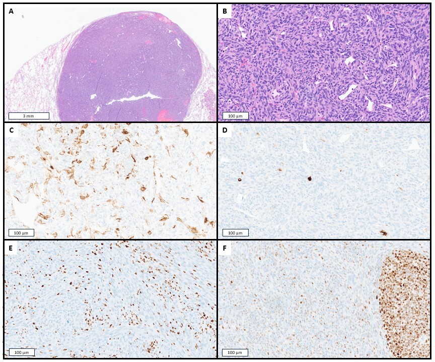
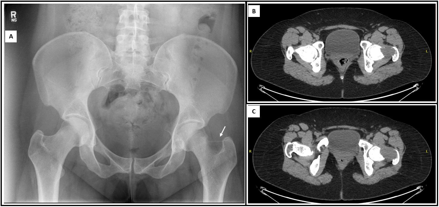
A 29-year-old female with no significant medical history presented with worsening pain in her left hip. Serial computed tomography (CT) scans revealed a growing lytic lesion in her left femoral neck (Figure 1). A CT-guided biopsy showed a malignant neoplasm growing within the bone. The tumour was composed of high-grade plump spindle cells arranged in a patternless fashion with occasional short fascicles. The neoplastic cells grew in association with thin endothelial-lined sinusoids that demonstrated a hemangiopericytoma-like vascular pattern. Mitotic activity was brisk, and punctate necrosis was identified. No osteoid, bone, or cartilage formation was present. Immunohistochemical (IHC) evaluation showed that tumour cells expressed vimentin and SATB2 with focal expression of HMB45 and smooth muscle actin (SMA). Definitive diagnosis was deferred to the excisional specimen.
The patient’s left proximal femur was resected, and histopathologic examination showed a malignant high grade spindle cell neoplasm (6.1 cm) centered in the medullary cavity of the epiphysis and metaphysis. The tumour was excised with negative margins (closest 0.5 cm), and it was negative for definite joint space or articular surface involvement. The morphologic and IHC findings were the same as those of the biopsy. The final diagnosis was ‘malignant spindle cell neoplasm most suggestive of malignant PEComa.’ Molecular ancillary studies provided no evidence of sarcoma gene fusions, including in TFE3 (SARCP 138 gene fusion panel, Mayo Clinic, Rochester MN).
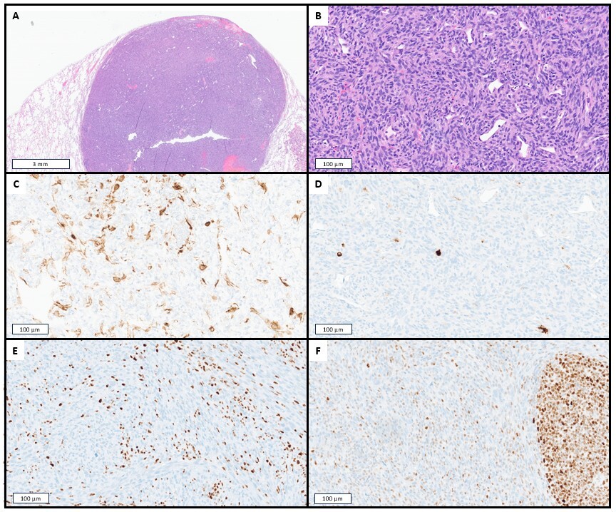
During postsurgical follow-up, two new suspicious lesions were detected on chest x-ray: one in the right upper lobe and one in the right lower lobe. These lesions had no correlate on a previous CT scan of the chest performed approximately four months prior. Wedge resections of both pulmonary lesions were performed, and histopathologic findings showed two metastatic deposits of PEComa (largest 1.7 cm). The metastatic lesions shared the same morphologic and IHC features as the bone primary (Figure 2). Both lesions were excised with negative margins. Additional ancillary testing was performed on one of the metastatic tumours. Programmed death ligand 1 (PD-L1) IHC was negative (22C3 clone). In-house clinical next-generation sequencing (NGS, Ampliseq Focus for Illumina Assay) did not identify any actionable variants, however several microsatellite sites were noted to be unstable, manifesting as small duplications and deletions at mononucleotide repeats. Subsequent IHC evaluation for MMR expression was performed on both the primary and metastatic tumours, revealing subtotal expression loss of MLH1 and PMS2 in both (Figure 2). MSI testing by polymerase chain reaction (Promega Corporation) found the tumour to be MSI-H. Testing for methylation of the MLH1 promoter was negative, raising the possibility of Lynch syndrome. Subsequent external comprehensive NGS (TruSight Oncology 500, Illumina) revealed a tumour mutation burden (TMB) of 16.5 mutations/Mb but was otherwise non-contributory, failing to identify mutations in MLH1, TSC1, or TSC2. The patient was referred to Medical Genetics and was found to be negative for Lynch syndrome, with no germline MLH1 mutation identified.
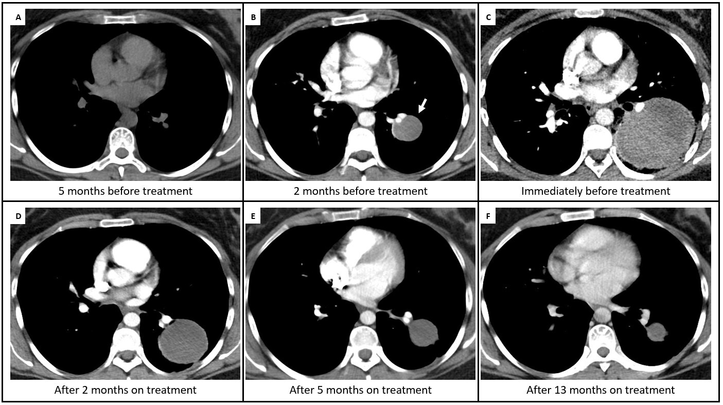
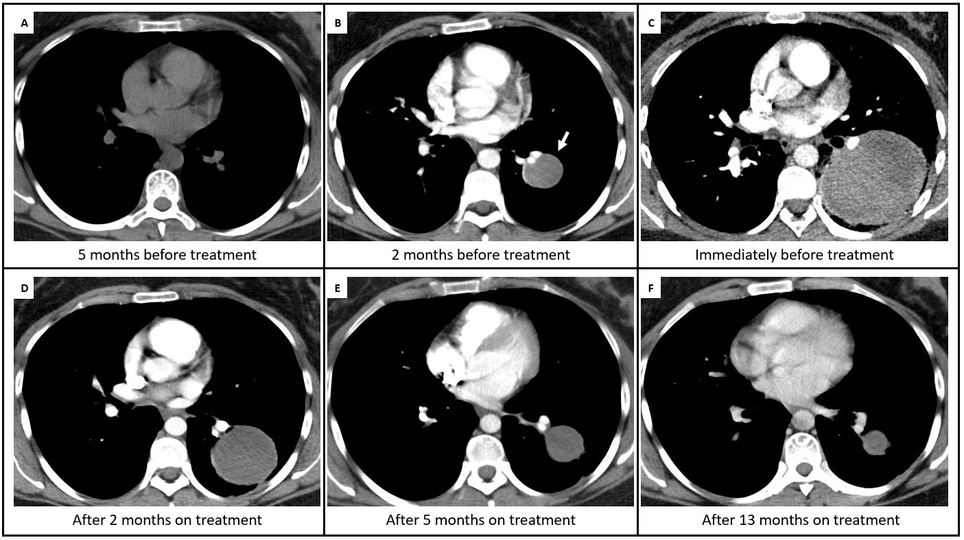
Subsequent CT imaging revealed a new rapidly enlarging lesion in her left lower lung lobe. At this time, the patient and care team elected to start systemic therapy rather than attempt to resect this new lesion. Approval for the use of single agent pembrolizumab was obtained from the Risk Committee at our institution. The patient received a baseline CT scan prior to beginning her treatment, showing rapid interval growth of her lung metastasis (to a maximum dimension of 6.5 cm). Approximately one week after her first dose of pembrolizumab, she developed small volume hemoptysis and was seen at her local emergency department. A CT scan of the chest with pulmonary embolus protocol was suspicious for rapid progression of the left lower lobe mass, which had increased in size to 8.0 cm. However, it was the opinion of her oncologist that this change represented pseudoprogression of her tumour. She continued with pembrolizumab and experienced no further major adverse effects. Subsequent CT scans over a period of 13 months showed a marked reduction in the size of the left lower lobe lesion (previously 6.5 cm, with reduction to 3.0 cm as of her most recent CT scan; Figure 3). At the time of writing (16 months since starting pembrolizumab), she is continuing to respond to her treatment with no major adverse events reported.
DISCUSSION
Our case shares several similarities with a small collection of published reports (Table 1). A recent report by Djerroudi et al. described a case of metastatic MSI-high PEComa in a 50-year-old man that arose in the context of Lynch syndrome, and was successfully treated with pembrolizumab.14 In that case, the patient’s known history of Lynch syndrome prompted the assessment for MMR protein expression. In our case, instability in the lengths of a few microsatellites was noted on initial NGS, prompting our decision to do follow-up MSI testing and MMR IHC. Of note, MMR IHC showed a subtotal loss of MLH1 expression in both our case and in the Djerroudi et al. case, whereas PMS2 expression had subtotal loss in our case versus complete loss in theirs.14 The lack of both pathogenic MLH1 mutation and MLH1 promoter hypermethylation in the tumour is surprising and provides no insight into the mechanism for greatly decreased MLH1 expression.
The TMB in our case (16.5 mutations/Mb) is comparable to that in the Djerroudi et al. case (21.2 mutations/Mb). Finally, PD-L1 expression was negative in both our case and theirs.14 This is in contrast to other reports that described the successful use of pembrolizumab in case reports of metastatic PEComa and epithelioid, all of which had high PD-L1 expression.15–17 MMR expression status was not reported in two of those cases and was intact in the third.
Clinical trials have shown that patients with locally advanced soft tissue sarcomas have a low rate of response to anti-PD-1 agents such as pembrolizumab. A number of the patients in these studies, however, experienced partial responses with periods of progression-free survival.18–20 The tumour MSI status of responders and non-responders in these studies is not reported.
MSI-high status has emerged as one of the most successful biomarkers in predicting the efficacy of immune checkpoint inhibitors in solid tumours.21 The KEYNOTE-158 study examined the pan-cancer efficacy of pembrolizumab in MSI-high tumours and included the enrollment of 14 patients with sarcomas, none of which were PEComas.22 Of those patients, the 12 that could be evaluated for tumour response ranged from having complete response (in one), partial response (in three), interval stability (in six), and progression (in two).22 These data collectively indicate that individual patients with advanced MSI-high sarcomas should be considered for anti PD-1 agents.
This case is the first to our knowledge to describe the successful use of immunotherapy for metastatic MSI-high PEComa arising in a patient without Lynch syndrome. Our report adds to a small but growing collection of evidence showing that metastatic PEComa can have strong and sustained responses to immunotherapy agents. This pharmacologic response appears to be related to tumoural PD-L1 expression in some cases and MSI in others. Further work is required to determine the best method for predicting response to anti-PD-1 agents in metastatic PEComa and other sarcomas. In the meantime, routine screening for MMRd in metastatic PEComa is recommended given the early evidence suggesting that it can be a predictor of response to immune checkpoint inhibition.