INTRODUCTION
Carcinomas are malignant neoplasms that arise from epithelial cells, which line the body’s surfaces, glands, and internal cavities. As the most widespread tissue type in the body, epithelial cells are highly susceptible to genetic mutations due to constant exposure to environmental toxins, mechanical stress, and cellular replication errors. This may explain why carcinomas account for approximately 80 to 90% of all human cancers.1
The American National Cancer Institute’s Surveillance, Epidemiology, and End Results (SEER) training modules2 broadly classify carcinomas into two major histological types:
-
Adenocarcinomas, which originate from glandular epithelial tissues and commonly occur in organs such as the colon, lungs, prostate, pancreas, stomach, breasts, and rectum.3
-
Squamous cell carcinomas, which arise from squamous epithelial cells and typically affect areas such as the skin, head and neck, esophagus, lungs, and cervix.4
Beyond these, other specialized subtypes include:
-
Urothelial carcinoma (formerly transitional cell carcinoma), arising from the urothelial lining of the urinary tract5;
-
Basal cell carcinoma, originating from basal epidermal cells of the skin6; and
-
Neuroendocrine carcinomas, derived from epithelial cells exhibiting neuroendocrine differentiation, found in organs like the lung and gastrointestinal tract.7
Each subtype differs not only in tissue of origin and morphology but also in molecular profile, biological behavior, and clinical prognosis.
Carcinomas are classified along a spectrum of differentiation, which reflects how closely tumour cells resemble their normal tissue counterparts in structure and function. This assessment is based on microscopic features such as cell shape, size, arrangement, organization, and the presence or absence of gland formation or keratinization.8,9
The differentiation spectrum includes:
-
Well-differentiated tumours, closely resembling normal tissue;
-
Moderately differentiated tumours, showing partial resemblance with notable abnormalities;
-
Poorly differentiated tumours, appearing more primitive and disorganized; and
-
Undifferentiated (anaplastic) tumours, lacking distinctive morphological and immunophenotypic features.10
Poorer levels of differentiation are typically associated with more aggressive clinical behavior and worse prognosis.11 Furthermore, undifferentiated tumours present diagnostic challenges due to their lack of specific histological features, often requiring extensive immunohistochemistry and molecular testing to exclude other entities. In these high-grade tumours, immunohistochemical patterns can be particularly unreliable due to phenotypic plasticity; they frequently exhibit a loss of typical site-specific markers (dedifferentiation) or the aberrant gain of unexpected markers. Such immunophenotypic instability, combined with their characteristic high metastatic potential, frequently obscures the identification of the primary tumour site. Clinical outcomes are generally poor.
Despite their clinical relevance, data on undifferentiated carcinomas remain limited. This retrospective study aims to address this gap by analyzing the clinical and pathological features of undifferentiated carcinoma patients diagnosed at Scarborough Health Network Hospitals over the past 23 years. We also present a detailed case report of a 39-year-old male with undifferentiated carcinoma of the colon, an exceptionally rare entity. Colorectal cancer is the third most commonly diagnosed malignancy globally12 and the second leading cause of cancer-related death.13 Through this analysis, we aim to contribute insights that may enhance current understanding and inform future diagnostic and therapeutic approaches.
METHODS
We conducted a retrospective study of patients diagnosed with undifferentiated carcinoma between 2002 and 2025 at Scarborough Health Network Hospitals. Patients were identified through a comprehensive search of the hospital’s pathology database.
Demographic variables collected included age at diagnosis and sex. Clinical variables included the primary tumour site, cancer stage at diagnosis, presence of metastasis, lymph node involvement, and lymphovascular invasion. All data were reviewed for accuracy by a designated researcher and pathologist supervisor.
RESULTS
A total of 13 patients met the inclusion criteria, including five males and eight females, with ages ranging from 39 to 81 years (see Table 1). The mean age at diagnosis was 57.8 ± 17.9 years for males and 67.9 ± 10.0 years for females. The primary tumour sites included five originating from the gastrointestinal tract (38.5%; colon, n=3; rectum, n=1; stomach, n=1), five from gynecologic organs (38.5%; ovaries, n=3; endometrium, n=2), two from the respiratory system (15.4%; lungs, n=2), and one from the genitourinary system (7.7%; ureter, n=1). Metastatic disease was documented in nine patients (69.2%), involving organs such as the brain, adrenal glands, omentum, mesentery, lungs, ovaries, bowel walls, and lymph nodes. Out of 13 cases, lymphovascular invasion was present in 6 patients (46.2%) while lymph node metastasis was present in 5 patients (38.5%), but notably, only 3 patients (23.1%) exhibited both.
Among the 13 cases in our retrospective cohort, which demonstrate the aggressive nature and diagnostic heterogeneity of undifferentiated carcinomas, the case of a 39-year-old male stands out as the most recent and diagnostically and molecularly complex. Unlike the other gastrointestinal cases, which followed a more conventional clinical pattern, this tumour exhibited a unique combination of rhabdoid morphology and atypical molecular alterations. This case represents an undifferentiated colon carcinoma, a rare and poorly characterized entity. Given the unusual histologic features and the limited data available in the literature, a detailed case report is presented below.
CASE REPORT
A 39-year-old South Asian male presented with a 4- to 5-month history of progressive anemia and constipation. He reported profound fatigue and unintentional weight loss exceeding 40 pounds. While awaiting an abdominal ultrasound, he was admitted to the hospital. He had recently been started on a proton pump inhibitor.
Initial lab investigations revealed a hemoglobin level of 40 g/L, which increased to 70 g/L following transfusions. An urgent esophagogastroduodenoscopy (EGD) and colonoscopy were performed. The EGD was unremarkable. Colonoscopy revealed a circumferential, ulcerated, ischemic-appearing lesion with a bleeding edge, starting at approximately 52 cm from the anal verge and extending down to approximately 45 cm from the anal verge. Multiple biopsies were obtained, and tattoos were placed proximal and distal to the lesion in the descending colon. Postoperative imaging confirmed a large lesion at the splenic flexure with no evidence of distant metastases. The patient’s symptoms of anemia, weight loss, and progressive constipation were attributed to blood loss from the ulcerated descending colon malignancy.
The patient returned to the hospital the evening prior to his scheduled surgery due to hematochezia and a drop-in hemoglobin from 90 g/L to 76 g/L. He received a unit of blood preoperatively and underwent an urgent laparoscopic anterior resection of the extended left colon. The hemicolectomy specimen was sent to pathology for further evaluation.
Gross Findings
The left hemicolectomy specimen measured approximately 33 cm in length with an average circumference of 9 cm. On the serosal surface, an area of puckering and induration was observed. Upon opening the bowel, a circumferential, necrotic, and friable tumour mass was identified in the splenic flexure, measuring approximately 13 × 11 × 2 cm. The mass involved the entire luminal circumference and was located at 6 cm from the proximal margin, 15 cm from the distal margin, and 3 cm from the closest mesenteric margin. No other discrete lesions were grossly identified.
Histologic Findings
Histologic examination of the left hemicolectomy specimen revealed a Grade 4 invasive undifferentiated carcinoma with a prominent sarcomatoid component (see Figure 1). The tumour was composed of rhabdoid cells with intracytoplasmic inclusions arranged in discohesive sheets and admixed with markedly pleomorphic giant cells. Extensive areas of necrosis were present.
The tumour involved the mucosa and invaded through the muscularis propria into the pericolorectal soft tissue. Lymphovascular invasion was identified in small vessels; however, no perineural invasion was observed. No precursor lesion or polyp of origin was identified. All resection margins were free of carcinoma, high-grade dysplasia, intramucosal adenocarcinoma, and adenomatous changes. All 25 examined lymph nodes were negative for metastatic carcinoma.
Immunohistochemical Findings
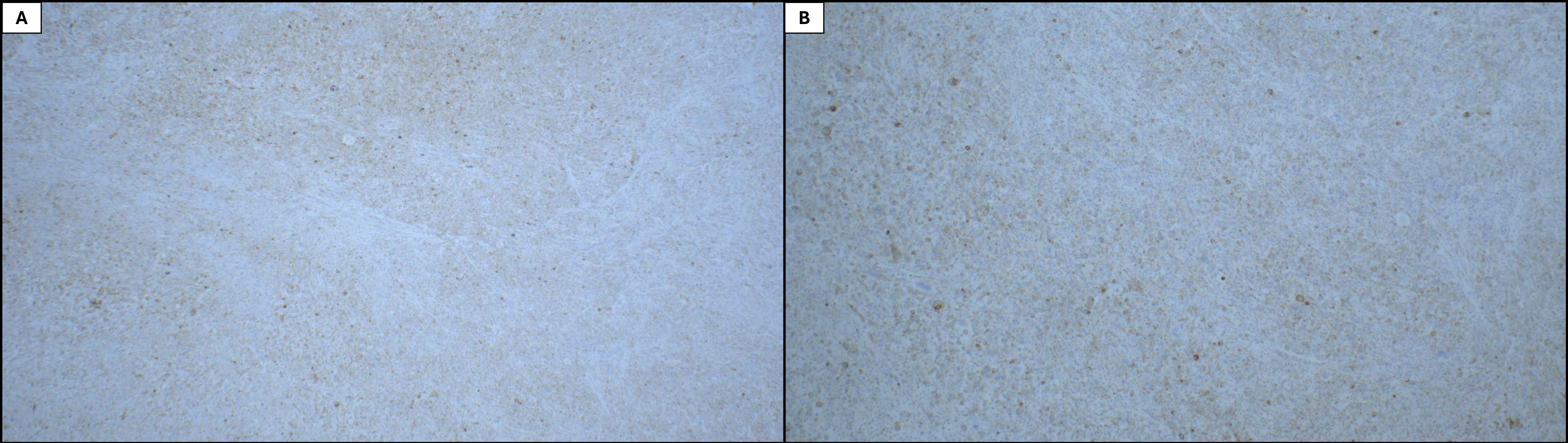
Immunohistochemical studies showed strong positivity for INI-1 (retained), pan-cytokeratin (AE1/AE3), and CK18. The tumour was negative for synaptophysin, chromogranin, CD56, CK20, CK7, desmin, actin, SMA, SMM, Melan-A, PAP, GATA3, NapsinA, TTF-1, HMB45, S100, ERG, SOX10, CDX2, PAX8, CD117, MDM2, CDK4, and ALK1. Notably, in-house synaptophysin immunostaining demonstrated scattered weak positivity. In contrast, repeat staining by an external consultant was negative, and upon their interpretation, the initial stain was also interpreted as negative (see Figure 2). There was equivocal, weak nuclear expression of SATB2 and DOG1 in only a subset of tumour cells.
Immunohistochemical studies additionally revealed retained nuclear expression of MLH1, PMS2, MSH2, and MSH6, which is highly correlated (>99%) with tumours that are microsatellite stable on molecular analysis.
Molecular testing revealed the following findings:
-
CKIT: Exon 17 D816Y mutation
-
BRAF: D594G mutation
-
PDGFRA: Wild type (no mutation detected)
-
RAS (KRAS and NRAS): Wild type (no mutation detected)
Based on these findings, the diagnosis was consistent with undifferentiated colorectal carcinoma, with a pathological stage of pT3pN0 as defined by the 8th edition of the American Joint Committee on Cancer Tumour Node Metastasis staging system.
Outcome
On postoperative day 5, contrast-enhanced computed tomography (CT) scans of the abdomen and pelvis revealed massive gastric distention requiring decompression, as well as expected postoperative findings, including small to moderate ascites, pneumoperitoneum, and extraperitoneal air. The study also demonstrated thickening and enhancement of small bowel loops, which could represent postoperative edema but might also reflect early ischemia. Other systems appeared unremarkable.
On postoperative day 183, a Persantine myocardial perfusion scan (Persantine MIBI) was performed to evaluate for myocardial ischemia due to suspected prior myocardial infarction and borderline Q waves on electrocardiogram (ECG). While the ECG portion of the stress test was nondiagnostic, nuclear imaging revealed a mild fixed perfusion defect in the mid to basal segments of the inferior myocardial wall with no evidence of reversible ischemia. Maintained wall motion and stable ejection fraction (70% rest, 71% stress) effectively ruled out active ischemia, indicating the defect was likely due to attenuation artifact rather than prior infarction.
On postoperative day 239 and again on day 397, contrast-enhanced CT of the chest, abdomen and pelvis demonstrated stable, unremarkable findings: no mediastinal, hilar, supraclavicular axillary, or pelvic lymphadenopathy; clear lungs and pleura without nodules or effusions; and preserved visceral organs, including an intact colonic anastomosis without masses, fluid collections, obstructions, ascites, or aggressive osseous lesions, with only incidental cholelithiasis noted on day 397. Endoscopic surveillance with digital rectal examination the following day revealed no significant abnormalities. Overall, there was no evidence of residual or recurrent malignant disease.
DISCUSSION
Undifferentiated carcinomas are exceedingly rare, accounting for only 0.2% of all carcinoma cases,14 yet their presence across multiple organ sites– as observed in our cohort– highlights their diverse origins and clinical significance despite limited representation in the literature. In this retrospective review, we identified only 13 cases of undifferentiated carcinomas between 2002 and 2025 at Scarborough Health Network hospitals. Primary locations in our series included the lungs, gastrointestinal tract (stomach, colon, rectum), and female reproductive organs (ovary and endometrium), and the urogenital tract (ureter). Patients ranged from 39 to 81 years old, with a slight female predominance (8 of 13 cases). A majority presented with advanced disease: many had lymph node involvement or distant metastases at diagnosis. These findings highlight the aggressive clinical course typical of undifferentiated carcinomas and the tendency for early dissemination.15 Identifying the site of origin can sometimes be challenging in this context, as undifferentiated tumours often lack organ-specific features and may present as metastatic disease of unknown primary.16 Despite the varied sites, the unifying theme in our cohort is the high-grade, anaplastic nature of the tumours and a generally poor prognosis associated with this diagnosis.
The case of the 39-year-old male with undifferentiated colorectal carcinoma is particularly noteworthy due to its rarity and diagnostic complexity. Undifferentiated colorectal carcinoma with rhabdoid features is infrequently reported in the medical literature, largely because the significant histopathologic heterogeneity of these tumours complicates their accurate diagnosis and classification.17 The presence of rhabdoid features further underscores the diagnostic challenge, as these features can overlap with various other malignancies and potentially confound the pathologic interpretation. Consequently, detailed immunohistochemical and molecular testing is crucial for distinguishing undifferentiated carcinomas from other aggressive tumours (such as melanomas, lymphomas, sarcomas, or high-grade neuroendocrine carcinomas), as this directly impacts prognosis and treatment planning. A diagnosis of undifferentiated carcinoma should only be made after all other possible entities have been carefully excluded.
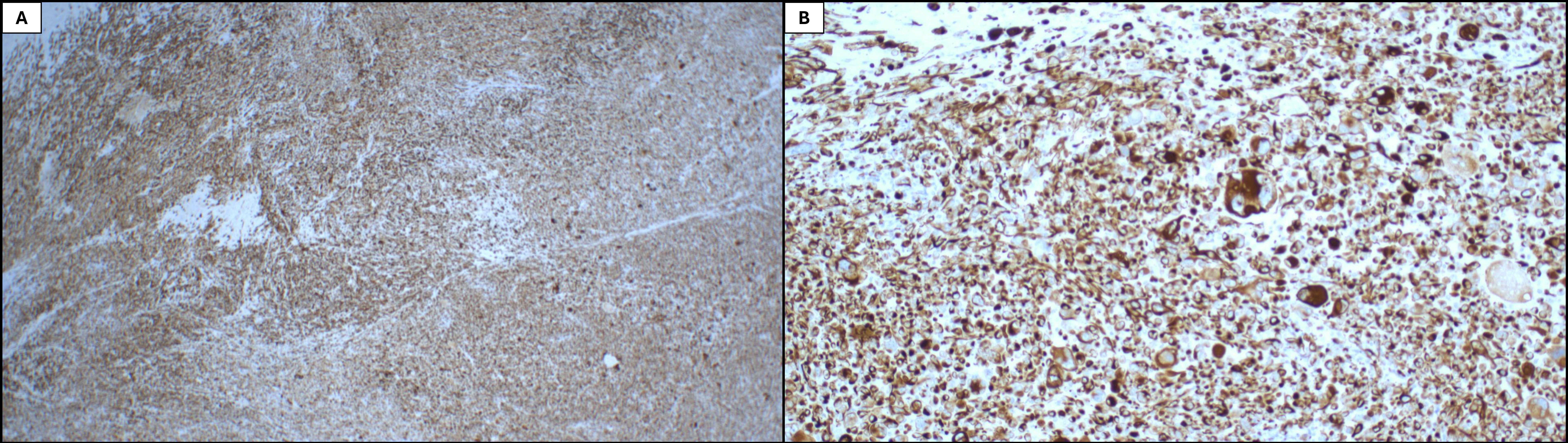
Morphologically, the tumour was a pleomorphic, high-grade malignant neoplasm lacking definitive features indicative of a specific line of differentiation. No adenomatous or other premalignant lesions were present in the background mucosa. Immunostains revealed that the tumour cells exhibited diffuse and strong immunoreactivity for the cytokeratins AE1/AE3 (see Figure 3) and CK18, definitively establishing an epithelial origin, as cytokeratins are markers of epithelial cells. Furthermore, a broad immunohistochemical panel was uniformly negative for other lineage-specific markers, including TTF-1 and Napsin A (lung), GATA3 (breast, bladder), PAP (prostate), and neuroendocrine markers (synaptophysin, chromogranin, CD56). Similarly, the tumour lacked expression of muscle-specific markers (desmin, actin, SMA) and melanocytic markers (S100, HMB-45, Melan-A, SOX10), effectively excluding these differential diagnoses.
The possibility of a germ cell tumour (GCT) was also specifically considered as part of the differential diagnosis as they are reported to account for up to 60% of all malignancies diagnosed in men between 20 and 40 years of age18; although the majority consist of testicular tumours, GCTs are also reported at extra-gonadal sites such as the retroperitoneum and mediastinum, originating from migrating primary germ cells that are left behind during fetal development.19,20 These tumours are a known source of high-grade malignancies that can display atypical imaging features and unusual growth patterns and may be mistaken for carcinomas if not specifically investigated.21–23 In our case, the identification of a CKIT (D816Y) mutation initially raised the suspicion of a GCT, as CKIT (the gene encoding CD117) is a characteristic driver in entities such as seminomas.24,25 However, the diagnosis was definitively excluded due to a striking discordance between the tumour’s genotype and its immunophenotype. Despite the presence of the mutation, the actual CD117 protein was not expressed, as confirmed by the negative immunohistochemical result. Furthermore, the tumour cells exhibited strong, diffuse expression of AE1/AE3 cytokeratins, a finding that effectively rules out these tumours, which are characteristically CD117-positive and AE1/AE3-negative.26,27
The identification of the CKIT (D816Y) mutation also necessitated the exclusion of a pleomorphic gastrointestinal stromal tumour (GIST), as approximately 80% of GISTs harbor KIT gene mutations that result in the constitutive activation of the KIT receptor and its downstream proliferative signaling pathways.28 While the majority of these mutations occur in the juxtamembrane domain encoded by exon 11, mutations within the activation loop of the kinase domain encoded by exon 17, such as the D816Y variant found in our case, are also common.29 Furthermore, our molecular analysis confirmed the tumour was wild-type for PDGFRA, a finding consistent with the mutually exclusivity of KIT and PDGFRA mutations in the oncogenic mechanisms in GISTs.28,30 Like in the case of GCTs above, immunohistochemical staining revealed a complete absence of the CD117 protein– which is usually strong and diffuse in GIST– despite the presence of a known activating KIT mutation.31 Crucially, DOG1 is considered a highly sensitive and specific marker for GIST regardless of CD117 expression28 or the underlying KIT/PDGFRA mutation.31 In our case, the identification of focal DOG1 immunoreactivity initially strengthened the consideration of a pleomorphic GIST; however, the weak and patchy nature of this staining, combined with the tumour’s morphology and diffuse immunoreactivity for two different keratins would argue strongly against this diagnosis.
Aside from DOG1, the immunohistochemical panel identified equivocal nuclear expression of SATB2. This transcription factor is used as a diagnostic marker for colorectal carcinomas,32 as current literature reports its positive expression in approximately 71% to 97% of primary and metastatic colorectal carcinomas.33 However, the reactivity in this case was limited to a weak, focal subset of tumour cells. Given the overlapping and non-specific staining patterns, molecular testing becomes essential, not only for supporting the tissue of origin in histologically ambiguous tumours but also for identifying actionable mutations. In colorectal cancer, mutations in genes like APC, TP53, KRAS, or PIK3CA are frequently observed34; their presence in an undifferentiated tumour supports a colorectal origin, even when morphology and immunohistochemistry are inconclusive.
Gene sequencing and exon-level copy number variant detection was performed by Next-Generation Sequencing on an Illumina MiSeq following library preparation using the TruSight Tumour 15 NGS kit (Illumina) to capture the regions of CKIT, PDGFRA, KRAS, NRAS, and BRAF with a minimum coverage equal to or greater than 500 reads. In addition to the previously noted CKIT (D816Y) variant, this analysis identified a concurrent BRAF D594G mutation, both of which are atypical for conventional colorectal carcinomas. The CKIT D816Y mutation has been previously associated with mastocytosis, acute myeloid leukemia, and testicular seminomas,35–40 while BRAF D594G has been most commonly reported in melanoma, diffuse large B-cell lymphoma, and non-small cell lung carcinoma.41–43 However, non-V600E BRAF mutations, such as D594G, are found in approximately 2% of colorectal cancer cases.44 One study on metastatic colorectal cancer identified ten patients with this mutant compared to 77 and 542 patients with BRAF V600E mutants and BRAF wild-type tumours, respectively.45 To date, no such research has been presented on the CKIT D816Y mutation. As previously explained, undifferentiated tumours often lose the molecular identity of their tissue of origin; as they become more genetically unstable, they can sporadically acquire mutations that are commonly seen in other cancers; this may have been the case for our patient’s CKIT D816Y mutation.
Moreover, this profile suggests potential targeted therapeutic opportunities that might be overlooked if treatment were based solely on histologic appearance. Notably, a reported case of colorectal carcinoma with a BRAF D594G mutation demonstrated a complete radiologic response within six months of treatment with a combination of the EGFR inhibitor cetuximab (Erbitux) and the MEK inhibitor trametinib (Mekinist).46
Ultimately, the final diagnosis was established through multidisciplinary correlation: although the immunohistochemical and molecular findings did not definitively establish the tissue of origin, they revealed features that were suggestive of, but not exclusive to, a carcinoma of colorectal origin; accordingly, radiologic–pathologic correlation was performed to ensure there were no alternative primaries or metastatic lesions. Following this, the tumour was managed as a primary colorectal carcinoma. Currently, no standardized treatment guidelines exist for these tumours, and prior studies have consistently associated them with poor clinical outcomes.47 Most colorectal cancer cases are managed primarily with surgical resection, especially when the tumour is localized.17 Moussaly and Atallah (2015) reported that most patients diagnosed with colorectal rhabdoid tumours who underwent surgical resection died within six months.48 Nonetheless, a small number of documented cases have shown patients achieving extended survival beyond one year.49–51 To the best of our knowledge, our patient had no evidence of residual tumour either macroscopically or microscopically. Due to clean margins and good response to surgery, we did not pursue targeted therapy at that time. Such a favorable outcome is particularly remarkable given the tumour’s exceptional rarity and its characteristically aggressive behavior.
In conclusion, this case reinforces the diagnostic complexity and unpredictability of undifferentiated carcinomas. It emphasizes the importance of a comprehensive approach, combining histopathological assessment, immunohistochemistry, and molecular profiling, and radiologic exclusion to arrive at an accurate diagnosis and guide appropriate treatment. As these tumours remain rare and poorly understood, continued documentation and study are essential for advancing clinical knowledge and improving care for future patients.
This manuscript was peer reviewed.
Acknowledgments
The authors would like to acknowledge Sabir Kazmi for providing technical support and assisting with the data collection process from the Scarborough Health Network database.
The authors declare that there are no undisclosed conflicts of interest regarding the publication of this paper. All authors have provided Clockwork Communications Inc. with non-exclusive rights to publish and otherwise deal with or make use of this article, and any photographs/images contained in it, in Canada and all other countries of the world.